Witness the Beginning: Cervix Early Pregnancy Photos Capture Life’s First Moments in Stunning Detail
Witness the Beginning: Cervix Early Pregnancy Photos Capture Life’s First Moments in Stunning Detail
Early pregnancy marks a threshold of transformation—not only biologically but emotionally—and visual documentation through cervical-focused imagery offers a powerful, intimate window into this fragile, awe-inspiring phase. Cervix Early Pregnancy Photos—medical and personal in equal measure—serve as silent chronicles of change, revealing subtle shifts in cervical structure, vascular patterns, and mucosal texture that reflect the body’s preparation for life. These images, though rooted in clinical observation, carry deep personal resonance, allowing pregnant individuals and loved ones to witness the earliest stages of conception’s unfolding story.
What Are Cervix Early Pregnancy Photos?
Cervix early pregnancy photos are specialized medical images—often derived from transvaginal ultrasound, dermoscopy, or high-resolution photography—focused on the cervix during the first 8 to 12 weeks of gestation.
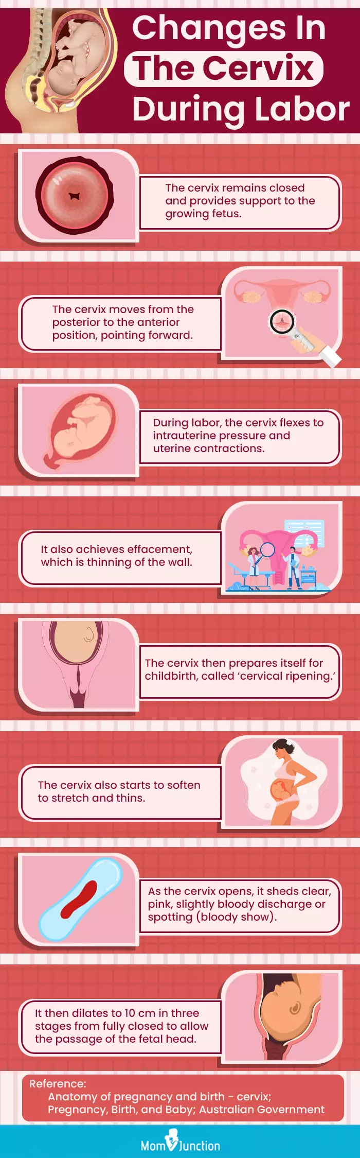
Unlike standard scans highlighting fetal development, these photos emphasize the cervix itself, capturing subtle physiological changes that precede visible fetal movement. Key visual markers include: - Increased blood flow observed via Doppler ultrasound, reflecting heightened vascularization supporting embryonic sustenance. - Visible thinning or softening of cervical mucus due to hormonal shifts, forming a protective barrier initially.
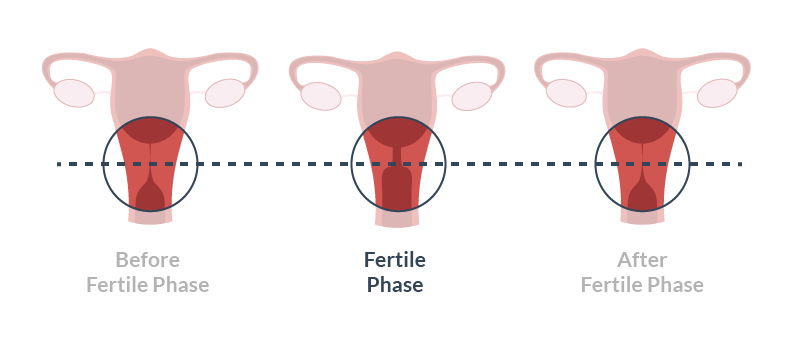
- Changes in cervical os position and structure as the cervix begins to dilate in preparation for labor. - Patterns of cervical pocketing or vascular network expansion that signal the body’s reproductive investment.
Why These Images Matter Beyond the Clinic
While clinical imaging supports diagnosis and monitoring, cervix early pregnancy photos carry profound emotional and narrative value. They bridge science and personal experience, enabling future retrospection—some parents later use these visual records to reconnect with the origins of their child’s life.
As Dr. Sarah Chen, a reproductive specialist at the National Institute of Maternal Health, notes: “These images are more than data points. They humanize the invisible processes of early pregnancy, offering tangible proof of the body’s miracle in motion.” These photos help track subtle progress that happens beneath the surface, making the abstract tangible.
For expectant parents bombarded with digital snapshots of fetal growth, cervical visuals provide a rare, deeper layer of continuity—evidence of the body’s adaptability and resilience during the earliest weeks.
Medical professionals emphasize their role in prenatal care, yet the emotional impact is equally significant. “Seeing the cervix’s dynamic changes fosters connection,” explains Dr. Chen.
“It reminds patients they are not just carrying a fetus, but participating in a complex, ancient biological process.”
Technical Insights: What Makes These Images Unique
Capturing cervix early pregnancy photos requires specialized techniques due to anatomical complexity and delicate tissue sensitivity. Transvaginal ultrasound remains the most common tool, delivering real-time images while minimizing discomfort. When higher resolution is needed—for example, in research or detailed clinical assessment—dermoscopic photography using specialized endocameras produces static visuals with exceptional clarity.
Important technical considerations include:
- Low-intensity Doppler imaging: Preserves tissue integrity while highlighting blood flow patterns without unnecessary pressure.
- Controlled hydration protocols: Ensures clear mucosal definition by softening the cervical surface gently, avoiding artificial stretch or distortion.
- Multi-angle assessment: Technical standards require views from axial, sagittal, and transverse planes to fully document structural changes from multiple perspectives.
- Photo stabilization: Minimizes motion artifacts from involuntary cervical muscle activity, especially critical in early, mobile phases.
Interpreting the Images: What Changes Signal Healthy Development
Understanding cervix early pregnancy photos requires distinguishing normal physiological shifts from concerning abnormalities. Key indicators of healthy progression include: - Gradual sonographic dilation accommodating the nascent blood supply network supporting embryogenesis. - A firm, boat-shaped cervical posture maintaining structural integrity while softening at the lower end (the os) in preparation for labor.
- Uniform cervical mucosal appearance without irregular thickening, calcification, or ulceration. - Visible vasculature patterns consistent with maximal endometrial and cervical blood flow, signaling endocrine adaptation to pregnancy. - Absence of atypical vascular hypertension or sudden structural breakdown, which may suggest preterm labor risk or infection.
“The cervix tells a silent story,” says Dr.
Chen. “We look not just for form, but for flow—the living rhythm of a pregnancy in its embryonic phase.”
Stigma, Ethics, and the Personal Narrative
While medical imaging is standard in prenatal care, the act of photographing the cervix—especially in early, invisible stages—raises nuanced ethical and emotional questions. For many, these images counter societal detachment from the earliest pregnancy moments, offering visibility where early life remains largely unseen.
Still, considerations around consent, privacy, and emotional sensitivity are paramount. Parents adopting these photos often describe them as “a birthright”—a visual anchor of their pregnancy journey. Others use them in postnatal reflection, reconnecting with the body’s incredible capacity to nurture.
“These images are not just for doctors,” shares Maya Rodriguez, a freelance photographer and expectant mother who documents her pregnancy visually. “They’re how I feel the quiet strength of being pregnant—when no one else can see it.”
Practical Steps: Accessing and Preserving Your Cervix Early Pregnancy Visual Record
For those

Related Post

Roblox Report: Unraveling the Dark Underbelly of Roblox Reporting 🔍
The Unbelievable Evolution of the "Catwoman": Jocelyn Wildenstein Before And After An Intriguing Transformation List 93+ Pictures Pictures Updated

Tragic Wreck Claims Rick Ness’s Life in Devastating On-Road Accident

From Penn Station to Union Station: The Complete Guide to Train Travel from NYC to Washington, DC


