What Does the Sylvian Fissure Separate? Unlocking the Brain’s Hidden Anatomical Divide
What Does the Sylvian Fissure Separate? Unlocking the Brain’s Hidden Anatomical Divide
Beneath the outer layer of the cerebral cortex lies one of the most critical yet often overlooked structures in human neuroanatomy: the Sylvian fissure. More than a mere groove or sulcus, this landmark separates two critical functional domains of the brain’s temporal lobe—separating auditory association from limbic processing. Its precise location and role hold profound implications for cognition, emotion, and neurological health.
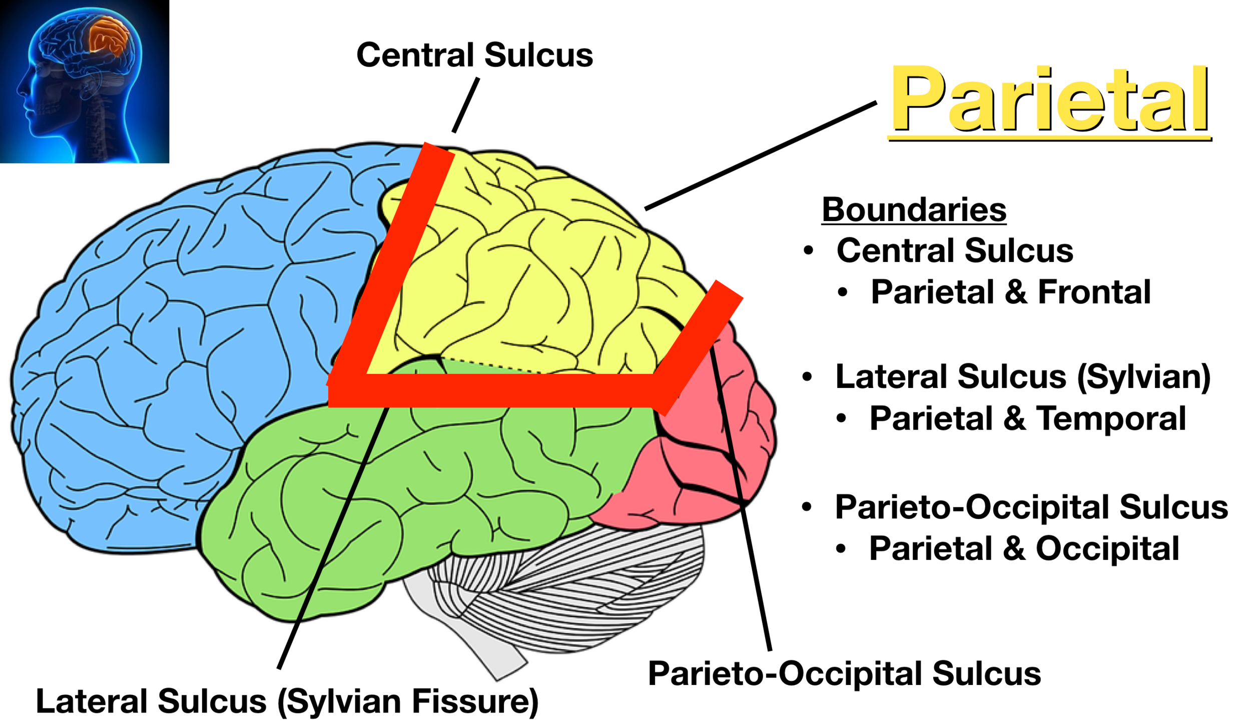
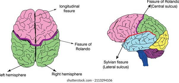
What exactly does the Sylvian fissure separate, and why does this distinction matter for understanding brain function and disease? The Sylvian fissure, also known as the lateral sulcus, cuts horizontally across the lateral surface of the brain, forming the boundary between the frontal, parietal, and temporal lobes. More specifically, it delineates the anterior temporal pole from the posterior inferior temporal regions, effectively separating auditory association areas—responsible for processing complex sounds—from limbic structures involved in memory, emotion, and social cognition.
This anatomical division is not merely structural; it reflects a deep functional segregation critical for how the brain integrates sensory input with emotional and mnemonic context. HbarTucked within the Sylvian fissure’s depth, the temporal lobe hosts key regions such as Wernicke’s area, pivotal for language comprehension, and the hippocampus’s surrounding circuits, essential for forming long-term memories. To fully grasp what the fissure separates, consider this: www.structural boundaries define not just physical segments but functional territories where neural networks specialize.
The fissure acts as a neural partition—one side dedicated to decoding complex auditory signals, including speech and environmental sounds, while the other side manages emotional resonance and autobiographical memory. This separation allows the brain to process auditory input with precision while simultaneously embedding it within a rich emotional and memory-based context. Claims about the Sylvian fissure’s role are grounded in decades of neuroanatomical research and modern imaging.
Advanced MRI techniques now visualize with remarkable clarity how this fissure tightens or broadens across individuals, correlating with variations in language ability, emotional regulation, and susceptibility to certain psychiatric conditions. For instance, studies show that individuals with dyslexia may exhibit subtle asymmetries in Sylvian fissure depth, potentially disrupting the balance between auditory processing and language comprehension. Similarly, abnormalities in fissure morphology have been observed in patients with schizophrenia, suggesting the fissure’s integrity influences the integration of sensory and emotional signals.
The Sylvian fissure’s anatomical precision reflects the brain’s evolution toward specialized yet interconnected processing. It does not merely demarcate lobes—it enables them to function with distinct roles while maintaining dynamic communication via the adjacent corpus callosum and associative tracts. This balance is vital: while unilateral damage to temporal regions may impair sound recognition or memory, the Sylvian fissure ensures that these functions remain compartmentalized yet collaborative.
Such compartmentalization allows for targeted interventions in cases of stroke or tumor, where surgeons must navigate around this landmark to avoid disrupting critical language or emotional pathways.
The Fissure’s Role in Cognitive and Emotional Integration
The Sylvian fissure’s division shapes how the brain synthesizes external stimuli with internal states. On one side, auditory cortex processes incoming sounds—recognizing phonemes, emotions in tone, environmental cues. On the other, limbic regions interpret these signals through the lens of memory and feeling, lending depth to language (“the warmth of a childhood laugh”) or contextualizing danger in a dropped noise.This dual processing explains why a seemingly neutral sound can evoke powerful emotional memories, a phenomenon rooted in the fissure’s precise anatomical orchestration of input and interpretation. Research also highlights the fissure’s influence on neuroplasticity. Its structural variation across individuals suggests adaptability in how the brain reallocates auditory and emotional tasks, especially after injury.
For example, in children with early hearing loss, compensatory neuroplastic changes may reshape the functional boundaries around the fissure to enhance visual or tactile processing. Such resilience underscores the fissure not as a static wall, but a dynamic boundary responsive to experience.
Clinical and Diagnostic Significance
In clinical neurology, the Sylvian fissure serves as a crucial landmark.MRI scans rely on its clear image to detect abnormalities such as tumors pressing into temporal regions, stroke impacting auditory pathways, or degenerative changes in Alzheimer’s disease affecting the hippocampus. Surgeons use its orientation to plan resections, ensuring critical language areas remain intact while removing pathological tissue. Neurologists and psychiatrists consider fissure integrity when evaluating conditions linked to temporal lobe dysfunction, from temporal lobe epilepsy—where seizure focus often lies near the fissure—to mood disorders influenced by limbic connectivity.
The fissure’s clear anatomical definition also aids in studying brain lateralization. While both hemispheres process auditory information, subtle differences in the Sylvian fissure’s curvature and depth between left and right sides correlate with specialized roles: the left’s modified fissure supports linguistic precision, while the right’s may enhance emotional prosody and spatial-auditory mapping. These asymmetries inform our understanding of how neural specialization develops and how imbalances contribute to disorders like autism or language delays.
The Future of Fissure Research: Imaging and Intervention
Emerging neuroimaging technologies—such as diffusion tensor imaging (DTI) and high-resolution 7T MRI—are refining our view of the Sylvian fissure’s microstructure. These tools reveal not just its gross anatomy, but subtle variations in white matter tracts linking adjacent lobes along its course. Computational modeling now simulates how disruptions in fissure geometry affect signal propagation between auditory and limbic systems, offering predictive insights into recovery after injury or during development.Therapeutically, targeting the fissure’s anatomical context may revolutionize treatments. Stimulation therapies, for instance, could precisely modulate activity in circuits near the fissure to improve language rehabilitation in stroke survivors. Gene and molecular studies are beginning to explore how genetic factors influence fissure formation and stability, with long-term goals of identifying biomarkers for early intervention in cognitive disorders.
In sum, the Sylvian fissure is far more than a visual marker on the brain’s surface—it is a vital anatomical and functional boundary, separating auditory processing from emotional and mnemonic integration in a way that underpins how we perceive, communicate, and experience the world. Every variation in its shape, depth, or orientation speaks to the brain’s intricate design and adaptive complexity. As neuroscience advances, the Sylvian fissure remains a crucial clue in decoding the neural basis of human cognition, emotion, and resilience—linking structure to story in the most intimate architecture of the mind.

Related Post

Master Language Skills with CoolMathGamesHangman: The Brain-Boosting Power of Interactive Vocabulary Practice
The Unseen Bonds: Decoding the Complex Relationship Between Jennifer Garner And James Garner

Unlock X Data Power: Sotwe.com Archive & Trends Search Powers Insightful Journalism

Brandon Call Net Worth: Tracing the Rising Star Behind Hollywood Charisma