Tracking the Invisible: Bacterial Colonies in a Petri Dish Unmask Microbial Secrets
Tracking the Invisible: Bacterial Colonies in a Petri Dish Unmask Microbial Secrets
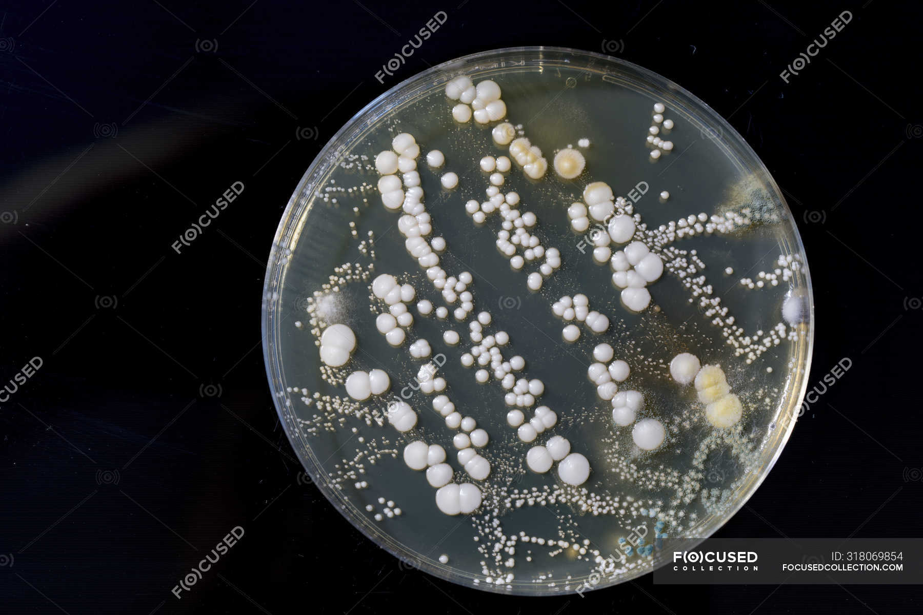
Observing bacterial colonies forming concentric rings on a nutrient-rich agar plate—visible evidence of growth, variation, and biology—these microscopic patterns reveal profound insights into microbial life. Bacterial colonies in a petri dish are far more than mere slides of bacteria; they serve as living laboratories for researchers, educators, and medical professionals to study growth dynamics, test antibiotics, understand genetic variation, and trace microbial evolution. Every colony tells a story encoded in DNA, making the simple act of culturing bacteria a cornerstone of modern microbiology.
A single Petri dish inoculated with a bacterial sample transforms into a dynamic microcosm. Under controlled conditions—temperature, humidity, and light—bacteria such as Escherichia coli or Staphylococcus aureus expand into visible colonies, each shaped by species-specific traits. These growth patterns are not random: colony morphology—size, shape, color, and surface texture—provides clues about the organism’s physiology and identity.
“Colonies are the voice of bacteria,” explains Dr. Elena Martínez, a microbiologist at the Institute of Applied Microbiological Research. “They quietly reveal how organisms respond to their environment, compete for resources, and interact within complex ecosystems.” The formation process begins with a small number of motile or spore-forming bacteria introduced onto a prepared agar medium.
As cells divide, they spread outward, consuming nutrients and oxygen while secreting waste products that alter the local chemistry. This spread creates clear, distinct colonies surrounded by a distinct zone where growth slows or halts—often due to nutrient depletion or toxic byproducts accumulating at the colony edge.
Understanding these patterns enables precise scientific applications.
Researchers use standardized methods like the Kirby-Bauer disk diffusion test—performed directly on agar plates—to assess bacterial resistance to antibiotics—a critical tool in the fight against antimicrobial resistance. Each zone of inhibition radiating from competing bacterial disks on the petri dish is a silent but powerful indicator of drug efficacy.
Beyond clinical labs, educational institutions harness Petri dish cultures to illustrate core biological principles. Students observe how isolated bacterial colonies from a grow pouch can grow into swarming, lysis-damaged, or pigmented colonies, each a physical manifestation of microbial diversity.“Watching from a slide, you touch the invisible world and witness its complexity,” notes Dr. Andrew Liu, a professor of microbiology at Stanford. “It transforms abstract concepts into tangible, visual data.”
Bacterial colony morphology varies significantly between species and even within strains, offering diagnostic power.
Coccus-shaped, round colonies of *Staphylococcus aureus* contrast sharply with rod-shaped, frequently clustered colonies of *Micrococcus luteus*. Variations may hint at metabolic capabilities—such as the ability to ferment glucose or produce pigments—directly informing identification under the microscope.
The incubation period—typically 16 to 48 hours—marks a waiting period that builds anticipation and precision. During this time, precise environmental control is essential: temperature fluctuations can delay growth or induce abnormal morphology, while contamination from airborne microbes risks invalidating results.Standardized protocols ensure reproducibility, letting scientists across the globe compare growth curves and colony characteristics reliably. Emerging technologies have elevated Petri dish analysis beyond visual inspection. High-resolution imaging, fluorescence staining, and automated colony counting now enable rapid quantification and deep phenotypic profiling.
“These tools unlock layers of data, from metabolic activity to gene expression patterns linked directly to colonial behavior,” says Dr. Priya Nair, a bioengineer specializing in microbial diagnostics. “It’s no longer just about counting colonies—each one becomes a data point in a vast digital microbiome database.” Yet, despite technological advances, the foundational act remains unchanged: a sterile bacterial sample introduced into agar, awaiting conditions that unlock its growth narrative.
Colonies emerge not just as living organisms but as mirrors—reflecting environmental pressures, genetic makeup, and biological interactions with precision honed over decades of scientific practice. In every petri dish, bacterial colonies serve as both witnesses and vessels of discovery. Their patterns carry information about infection mechanisms, evolutionary adaptation, and ecological roles.
They bridge the gap between theoretical microbiology and real-world application, grounding abstract science in observable, measurable reality. As we continue to explore microbial frontiers—from antibiotic development to probiotic therapies— Bacterial colonies in agar remain the quiet architects of knowledge, one visible ring at a time.


Related Post

Everything You Need To Know About Jonathan Stoddard's Wife

Exploring The Life Of Salena Zito: Her Husband And Family Dynamics


