The Hidden Guardian: Unlocking the Medical Significance of the Petrous Part of the Temporal Bone
The Hidden Guardian: Unlocking the Medical Significance of the Petrous Part of the Temporal Bone
Beneath the skull’s intricate architecture lies a small but supremely vital structure—the petrous part of the temporal bone—whose profound anatomical complexity underpins critical neurological, auditory, and facial functions. Often overlooked in introductory anatomy, this fuse-shaped segment harbors sensory organs, cranial nerves, and vascular pathways essential for balance, hearing, and facial expression. Its layered composition and deep-seated location make it both a cornerstone of cranial neuroanatomy and a frequent focus in clinical diagnostics.
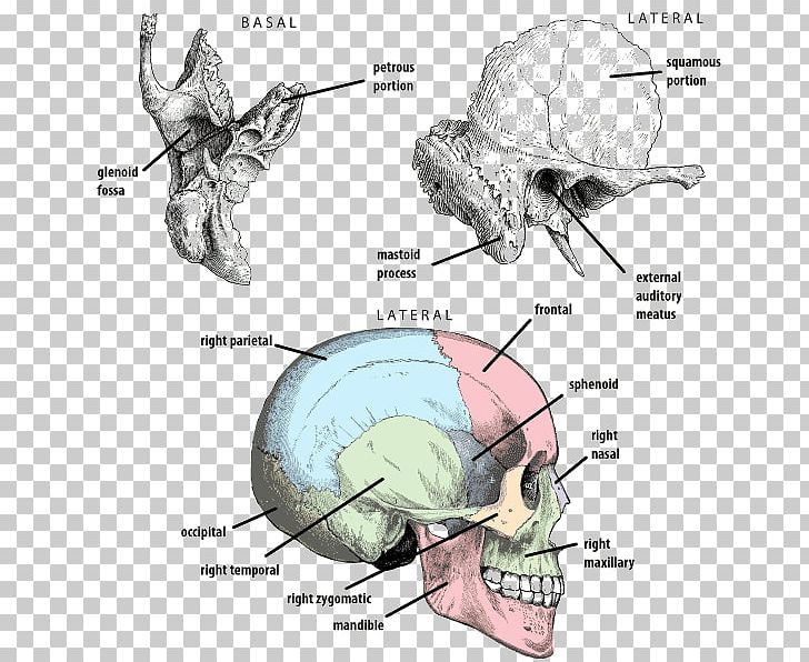
Comprising less than one percent of the temporal bone’s total volume, the petrous segment extends from the base of the temporal bone to the foramen magnum, housing the inner ear’s bony labyrinth and branching cranial nerves.
*“This tiny bone is a fortress of biological necessity,”* notes Dr. Elena Vasquez, an otolaryngologist specializing in skull base pathologies. *“Its role in perception and movement cannot be overstated—despite its diminutive size, it holds some of the most sensitive neural circuits in the human body.”*
Anatomy Unveiled: Layers and Landmarks of the Petrous Bone
The petrous portion is defined by its dense, lime-carbonate mineral structure, optimized to protect delicate neural and vascular elements.
It consists of several overlapping regions: the otic capsule enclosing the cochlea and semicircular canals, the internal auditory canal (IAC) housing cranial nerves VII, VIII, and laterally the trigeminal nerve (V3), and lateral facetal projections extending toward the jugular foramen.
- Otic Capsule: Encases the inner ear—perilymph-filled sac containing the cochlea (sensity hearing) and vestibular apparatus (balance). Its thick osseous wall dampens external pressure fluctuations.
- Internal Auditory Canal (IAC): A narrow 질 поías通故障 in the petrous bone, transmitting cranial nerves VII (facial), VIII (vestibulocochlear), and V (trigeminal, with its third division), responsible for taste, hearing, balance, and facial motility.
- Foramina: Seven key passageways—superior orbital fissure, internal acoustic meatus, jugular foramen, and internal carotid canal—all critical for neurovascular communication.
- Facial Convexity: The petrous region’s outer wall curves inward, protecting central neural circuits while allowing passage of neurovascular structures.
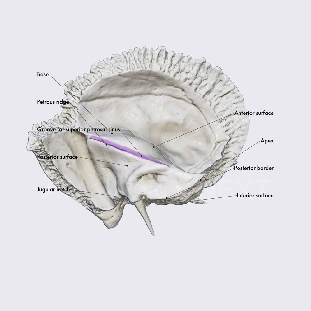
Visually, the petrous bone occupies the base of the temporal fossa, forming the skull base’s most posterior and inferior portion. Its basal surface articulates with the occipital bone at the foramen magnum, while its apical tip contributes to the skull’s overall structural integrity, anchoring the temporal ossicle’s cryptic yet crucial functions.
Critical Structures Enclosed: Sensory and Motor Pathways
Encoded within the petrous bone’s limestone labyrinth are life-signaling nerve bundles and fluid-filled spaces, making it a nerve center of extraordinary complexity.
The internal auditory canal serves as a transm練には altogether vital role, with cranial nerve VIII (vestibulocochlear) branching symmetrically to transmit auditory and equilibrium cues. Damage here—whether from nozzeikeal tumors, trauma, or infection—can permanently impair balance or induce deafness, underscoring the region’s


Related Post
Alicia Richards ABC27 Bio Age Height Education Husband Salary and Net Worth

Ping Pong Player: The Next Evolution of Smart Table Tennis Technology
Burnt Toast Has a Strong One: This Woman’s Terrifying Journey Awakens the Uneasy Truth About Trauma, Memory, and Silence

The Rise of Realistic Online Basketball Games: Where Practice Meets Immersion