The Elevated Ridges of the Brain Are Called the __
The Elevated Ridges of the Brain Are Called the __
Beneath the sleek, wrinkled surface of the human brain lies a hidden topography of functional significance—elevated ridges alternately crowned by gray and white matter, forming a network of profound neurological importance. These structures, known as the __, serve as anatomical pillars and functional hubs, orchestrating cognition, emotion, and identity. More than mere contours, the brain’s elevated ridges represent the brain’s most sophisticated architecture, where critical neural circuits converge, enabling human thought, perception, and behavior.
The Structural Identity: What Are These Elevated Ridges?
The elevated ridges of the brain are formally referred to as the **cornu gyri**—though commonly sinddied as the **corpus callosum’s prominent ridges** and especially recognized as the **central gyri** and **lateral gyri** within the broader neocortical landscape. However, collectively, these elevated, ridge-like formations are most precisely termed the **cortical ridges**, with the most prominent and functionally significant ridges being the **longitudinal folds** and the **gyral crests** that demarcate key lobes. The cerebral cortex is not a smooth canvas but a billboard of folded ridges and grooves—gyri (ridges) and sulci (grooves)—creating a surface area expansion by up to tenfold within the constrained vault of the skull.This folding increases synaptic connectivity and processing capacity, a biological marvel that underpins advanced cognition. The most definitive elevation among these is the **longitudinal fissure**, the deep groove separating the cerebral hemispheres, flanked by subtler but vital ridges like the **precuneus gyrus** and **parietal longitudinal ridges**. десяActivity of these ridges extends beyond anatomy—they are dynamic forums for neural integration.
The **corpus callosum**, though not a gyrus, is fringed by critical ridges that enable corpus-wide communication, effectively linking the left and right hemispheres in a seamless exchange of sensory, motor, and cognitive data. Meanwhile, the **inferior longitudinal gyrus** along the temporal lobe supports complex visual processing and memory formation, anchoring perception in spatial and associative networks.
Functional Significance: How These Ridges Shape Cognition
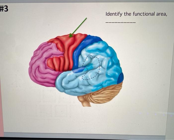
The elevated ridges are not passive landmarks but active centers of high-level brain function.Each ridge corresponds to a specialized region tuned to distinct neural tasks. For example, the **dorsal longitudinal gyrus** contributes to spatial reasoning and navigation, while the **ventral occipitotemporal ridges** support object and face recognition—critical for social interaction. These ridges house densely packed **neuronal cell bodies, dendrites, and synaptic junctions**, creating microscale command centers where information is synthesized.
Research using high-resolution fMRI and diffusion tensor imaging reveals that activity across these ridges synchronizes across brain networks, facilitating tasks from language comprehension to decision-making and emotional regulation.
“The cortex’s folded architecture turns distance into density,”* declares Dr. Elena Moreau, neuroanatomist at the Max Planck Institute for Brain Research.Neuroplasticity studies further demonstrate that stimulation or damage to specific ridges alters function predictably. For instance, injury near the **inferior frontal gyrus** impairs language production, whereas modulation of the **superior temporal gyrus** affects auditory processing and auditory hallucinations in schizophrenia. This functional localization underscores the ridges’ indispensable role in maintaining mental equilibrium.*“The elevated ridges are not just folds—they are hubs where perception becomes thought, and sensation becomes experience.”
Development and Evolution: Why the Ridges Matter Across Lifespan
From prenatal development onward, cortical folding begins in late gestation, guided by genetic blueprints and mechanical pressures within the skull. The timing and pattern of ridge formation are tightly regulated—delays or disruptions correlate with neurodevelopmental disorders such as lissencephaly, where reduced folding impairs cognitive development. > “The evolution of deep cortical ridges marks a key threshold in primate intelligence,” notes Dr.James Wu, a comparative neuroanatomist. “Humans exhibit more pronounced gyral complexity than other mammals, enabling richer language, abstract reasoning, and social complexity.” Beyond development, these ridges reflect lifespan adaptation. In aging brains, progressive thinning of cortical layers increases ridge visibility on scans, sometimes compensating for neuronal loss.
Yet, pathological folding changes—such as in frontotemporal dementia—disrupt ridge integrity, accelerating functional decline.
Clinical and Technological Frontiers: Mapping the Brain’s Gyral Landscape
Modern neuroimaging technologies now allow detailed 3D mapping of these elevated structures, transforming neuroscience and medicine. Preoperative MRI registering cortical folds enables precise localization for epilepsy surgery, minimizing damage to functional tissue.Advanced techniques like tractography visualize ridges’ white matter tracts, revealing connectivity patterns invisible to conventional imaging. In brain-computer interface (BCI) research, identifying ridge architecture improves electrode placement accuracy, enhancing signal decoding for paralysis patients and neural prosthetics. Moreover, deep learning models analyzing gyral morphology offer early biomarkers for disorders like autism and Alzheimer’s, shifting diagnosis from symptoms to structural signatures.
Future Directions: Beyond Structure Toward Dynamic Function
As neuroscience advances, the study of the elevated ridges is shifting from static anatomy to dynamic function. Emerging tools like optogenetics and real-time neural recording are decoding how ridge-level circuits support predictive coding, consciousness, and creative cognition. Researchers now explore whether targeted stimulation of specific gyri can modulate pain perception, mood, or cognitive load—ushered in a new era of personalized neurotherapeutics.Understanding the brain’s elevated ridges thus goes beyond labeling tissue—it unlocks insight into what makes human thought unique. These ridges, once mere anatomical curiosity, are now central to decoding the neural basis of mind. The elevated ridges of the brain, collectively known as the **cortical gyri**—and particularly prominent zones like the **longitudinal and lateral gyri**—represent the brain’s most intricate architecture.
Far from passive folds, they are dynamic centers of integration, enabling perception, language, emotion, and higher cognition. From prenatal development to aging, their structure defines functional capacity, making them indispensable to human identity and resilience. As technology and neuroscience converge, mapping these ridges deepens our grasp of the mind’s physical foundation—evidence that mind and matter are inextricably linked in the folds of the cerebral cortex.

Related Post
Henry Santos: The Soulful Voice Unveiled — Age, Legacy, and Global Impact
Jeffrey Gundlach Doubleline Bio Wiki Age Height Wife Cnbc Predictions and Net Worth
Unlocking Pigs’ TactiliciousWorld: How Sensory Papillae Shape Pig Skin Experience
Unveiling Benjamin Levy Aguilar Wife: Her Life Story