Medial Definition Anatomy: The Master Map of the Human Body
Dane Ashton
1579 views
Medial Definition Anatomy: The Master Map of the Human Body
At its core, medial definition anatomy refers to the precise anatomical positioning and structural relationships of body parts relative to the midline—or "medial" as defined in medical terminology—serving as a foundational framework for clinical diagnosis, surgical planning, and medical education. By anchoring every organ, muscle, bone, and nerve to a central midline reference, this anatomical system ensures unambiguous communication across disciplines and enables accurate visualization of complex human systems. As the Body Taxonomy Initiative acknowledged, "Medial definitions provide the objective coordinate system upon which all spatial medicine is built," transforming subjective descriptions into tangible, measurable data essential for precision healthcare.
Understanding medial anatomical references begins with the basic concept of the median plane—a vertical plane that divides the body into equal left and right halves, running from the crown of the head down to the pelvis. This central axis does more than classify symmetry; it defines key landmarks such as the mid-sternal line in thoracic anatomy or the midline of the intervertebral spaces in the spine. These reference points become critical for locating structures during physical exams, imaging, and procedural interventions.
For example, when surgeons speak of a lesion “medial to the aorta,” or pain “permedial to the tibial nerve,” they are invoking a spatially accurate, context-rich description rooted in standard anatomical language.
Core Components of Medial Anatomy: Symmetry, Mirroring, and Functional Alignment
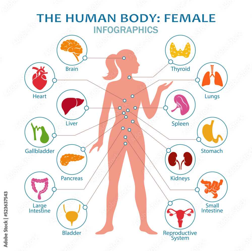
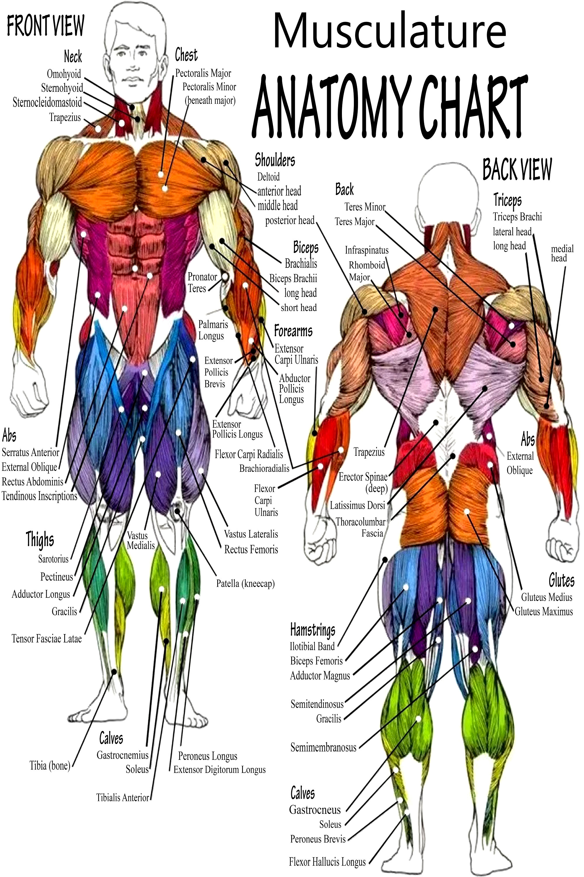
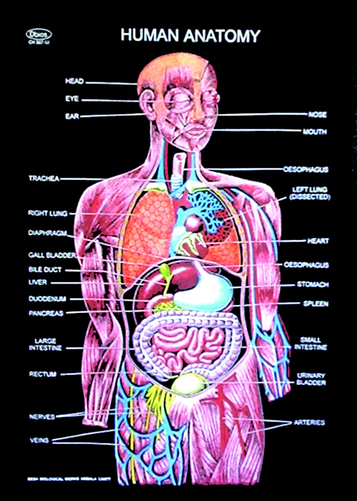
The human body operates on principles of bilateral symmetry, with major systems arranged in mirror-like patterns relative to the midline. Organs such as the heart and liver lie predominantly along the medial axis, though subtle variations—like slight differences in cardiac chamber positioning—highlight the limits of perfect symmetry. This mirroring extends to musculoskeletal structures: the biceps brachii and triceps reside symmetrically, yet their proximity to the median plane facilitates coordinated movement and force distribution.
Another defining feature is the functional alignment of anatomical structures. Tendons, ligaments, and fascial sheaths extend from one side of the body to the opposite, reinforcing mechanical efficiency through balanced tension. Consider the iliopsoas muscle, whose origin on the lumbar spine’s medial surfaces transitions into insertion along the femoral head—this medial trajectory aids in hip flexion while maintaining core stability.
Such alignment is not coincidental; it reflects evolutionary optimization, ensuring proximity between opposing anatomical systems enhances responsiveness and resilience. As anatomical scholar Trevor A. Griffin notes, "Medial relationships are not just positional—they’re dynamic, shaping how tissues interact under stress and movement.”
Critical Medial Landmarks in Clinical Practice
In clinical settings, medial anatomical definitions guide everything from diagnostic imaging interpretation to surgical incisions.
Radiologists rely on positional accuracy, identifying structures “medial to the trachea” or “lateromedially to the spinal cord” to avoid confusion in complex scans. Surgeons use medial references to navigate confined spaces—like placing a catheter “medial to the carotid artery” to prevent vascular injury—or during spinal procedures where “medial to the facet joint” indicates safe instrument placement.
Spinal Anatomy: Medial deviations such as medial spinal curvature anomalies (e.g., dorsolateral scoliosis) are tracked using mid-line imaging to assess progression and treatment needs.
Neuroanatomy: The corpus callosum’s midline positioning is essential for understanding interhemispheric communication; lesions “displaced lateromedially” disrupt neural integration.
Orthopedics: Medial epicondyle of the humerus serves as a palpable landmark for elbow joint evaluations and injury localization in repetitive stress cases.
The importance of medial precision is especially evident in minimally invasive procedures, where virtual guides map anatomical corridors defined by midline reference points.
A laparoscopic surgeon inspecting the pelvic cavity may identify “medial to the pubic symphysis” to safely dissect adnexal structures, reducing risk through spatially informed decisions. In this sense, medial anatomy transforms abstract coordinates into actionable blueprints.