From Cell Division’s Core: A Visual Journey Through the Phases of Mitosis—Portraits of Cell Division in Action
From Cell Division’s Core: A Visual Journey Through the Phases of Mitosis—Portraits of Cell Division in Action
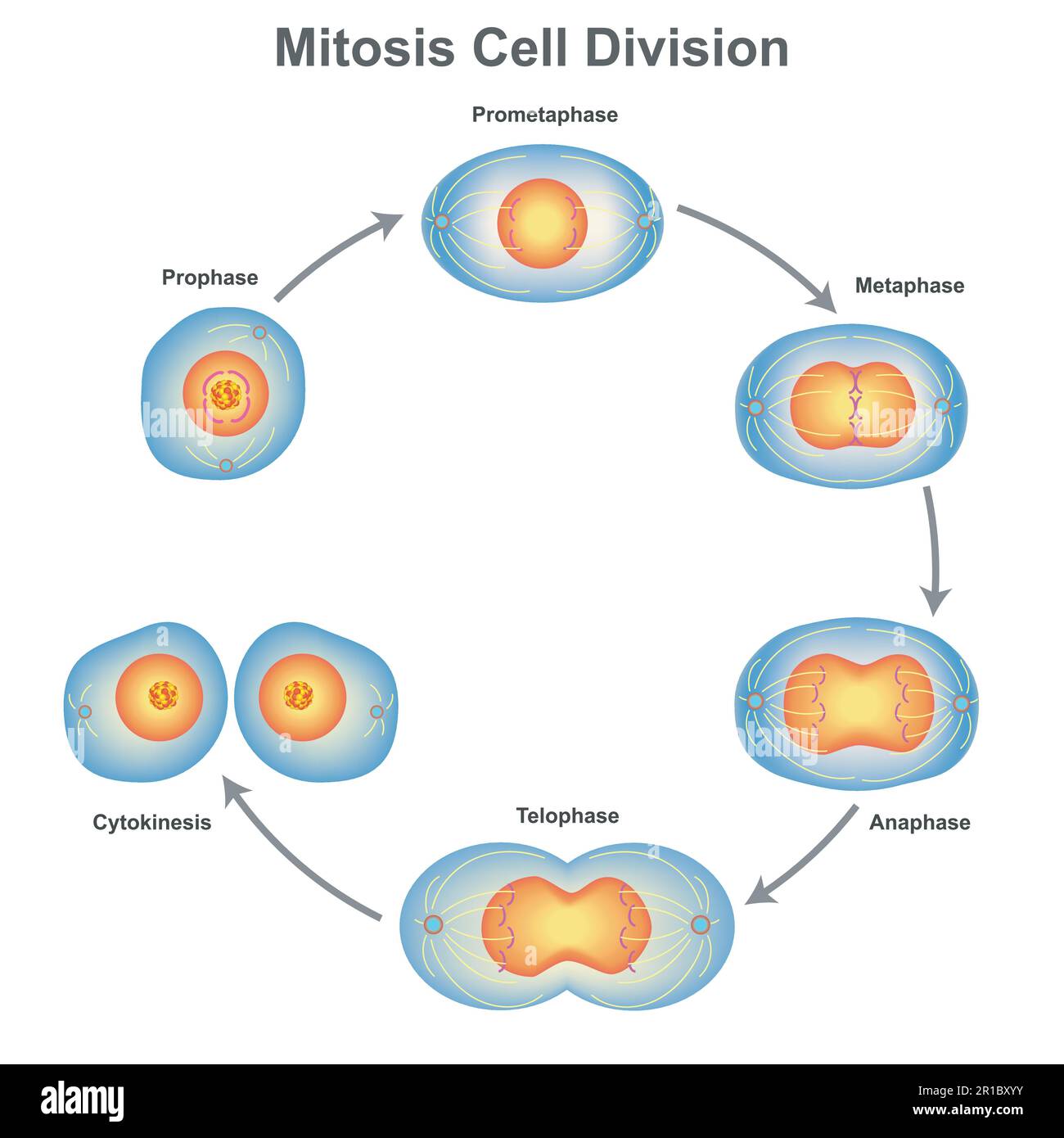
Mitosis, the precise mechanism of eukaryotic cell division, ensures each daughter cell receives an identical, complete set of chromosomes. This process is a masterclass in cellular precision—dividing the genome with unwavering accuracy across every living cell. With the aid of modern microscopy and vivid imagery, the four distinct stages of mitosis unfold like a dramatic sequence of microscopic theater: prophase, metaphase, anaphase, and telophase.
High-resolution pictures of each phase reveal a choreography of protein complexes, spindle microtubules, and chromosome dynamics clearly illustrating how life’s blueprint is faithfully copied and distributed. Understanding mitosis is fundamental to biology—grounding insights in genetics, cancer research, developmental biology, and regenerative medicine. Every stage represents a delicate phase where molecular machinery assembles to achieve equitable cell division.
Capturing these moments through detailed images transforms abstract concepts into tangible visual narratives, enabling scientists and students alike to grasp the mechanics of division with unmatched clarity.
During
The First Act: Prophase—Chromosomes Align for Fate
, intricate reorganization begins within the nucleus. As chromatin fibres condense into distinguishable chromosomes, each composed of two sister chromatids joined at the centromere, the nuclear envelope begins to fragment.Microtubule-sparing proteins disassemble, freeing motor proteins and kinesins. By electron microscopy, thousands of condensed chromosomes weave through the nuclear space, poised to migrate. Photographs of prophase reveal striking contrast—dense, linear structures moving toward opposite poles, marking the onset of division.
“Seeing chromosomes condense is like watching the cell’s DNA prepare for its ultimate split,” notes Dr. Elena Ruiz, molecular biologist at Stanford University. Prophase effectively launches the division sequence, ensuring genomic integrity before sister chromatids are unleashed.
In
Metaphase: The Equatorial Balance—A Celestial Alignment
, chromosomes orchestrate a breathtaking symphony at the cell’s equator. Under high-magnification imaging, the mitotic spindle—a lattice of microtubules—anchors each chromosome precisely at the metaphase plate, centering the paired chromatids like planets orbiting a star. This alignment is no accident: roles struck by motor proteins, spindle microtubules tugging from opposite poles, create tension that confirms accuracy.“The metaphase checkpoint ensures no cell proceeds until all chromosomes are properly aligned—a quality control step vital for genomic stability,” explains Dr. Marcus Tafari, expert in cell cycle regulation. Dynamic images capturing this balance emphasize symmetry and coordination—key to preventing division errors linked to aneuploidy, a signature of many cancers.
As
Anaphase Ignites: Sister Chromatids Part—The Physics of Division
begins, the tension shifts. The centromere splits, releasing sister chromatids that are rapidly pulled apart by shortening kinesin- and dynein-driven microtubules. Emerging pictures reveal a stunning ballet: chromatin fragments racing toward opposite poles, spindle microtubules shrinking like threads under load, and the cleavage furrow deepening as cytokinesis prepares.“Anaphase is where cell division truly cleaves—the moment sister chromatids separate with relentless force,” remarks Dr. Linneaにも, cellular dynamics specialist. Time-lapse photographs track their trajectory in real time, exposing the speed and precision: chromosomes travel up to hundreds of micrometers in minutes.
This phase is critical—missteps here produce unequal inheritance, a disruptor in development and disease.
Completing the cycle,
Telophase Restores Order—Nuclei Reassemble and Karyoplasts Return
marks the final retreat from chaos. As chromosomes decondense near the poles, nuclear envelopes reform around each set, reestablishing protective boundaries.The vacuole-rich cytoplasm concentrates around them, and the mitotic spindle disassembles. Electron micrographs show neat nuclear membranes resealing, chromosomes expanding into chromatin and doubling in size before nuclear envelope reassembly. “The transition from highly condensed to decondensed chromatin exemplifies cellular renewal,” notes Dr.
Ruiz. When viewed across all phases, mitochondrial images paint mitosis not as a mechanical process, but as a timeless dance of architecture, force, and molecular synchrony—essential for life’s continuity. The intricate choreography captured in mitotic phase pictures reveals more than cellular mechanics; it underscores the fragility and precision intrinsic to every living cell.
Each photograph tells a story of survival—coordination that persists across billions of divisions—and illuminates pathways to understanding disease and advancing regenerative therapies. From prophase condensation to telophase reintegration, the visual documentation of mitosis remains indispensable in modern biological science.


Related Post
Harrisburg Garage Sales Steal These Deals Before Everyone Else Does

20 Mg in Grams: Decoding a Critical Metric Across Industries and Everyday Life

Abbey Phillip: Redefining Innovation Through Purpose-Driven Impact

Springfield, Ohio City Council: Unpacking Party Affiliations and Their Political Impact